Home
Product
List
-
-
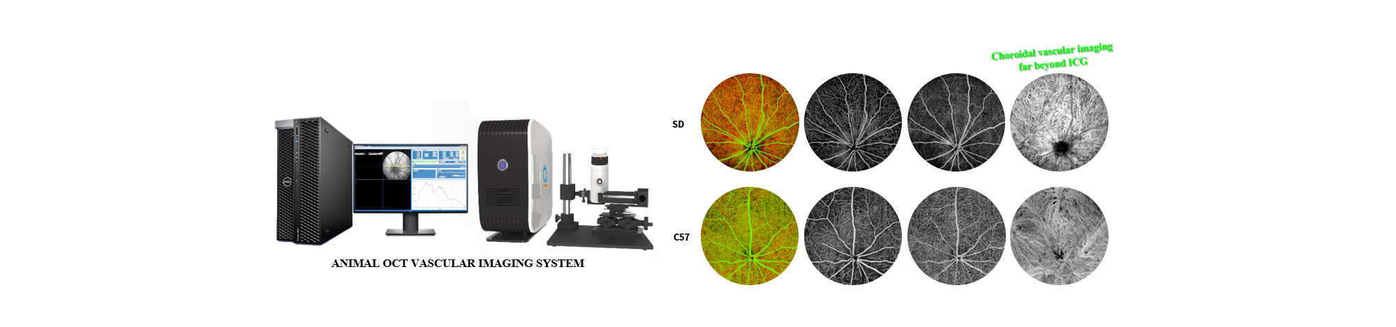
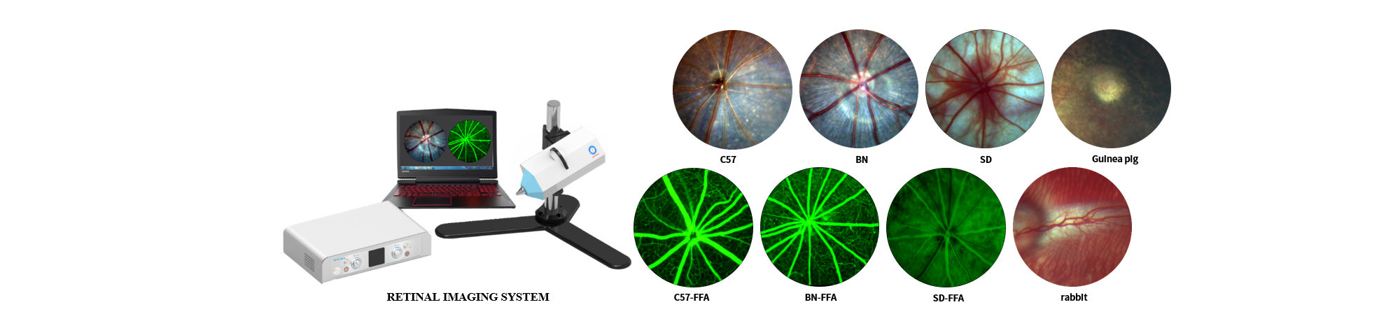
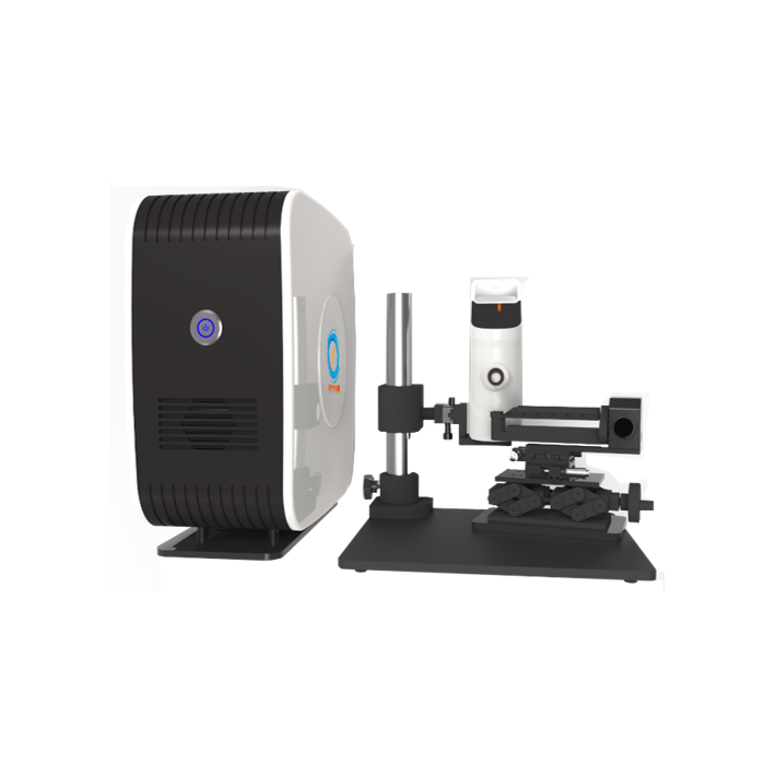
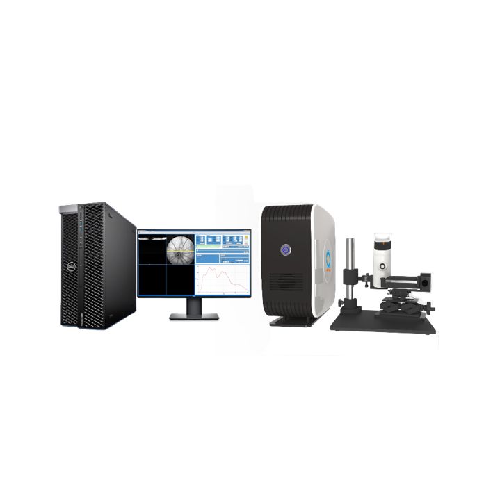
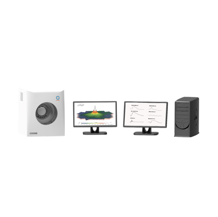
It is used for the research of disease screening, pathology, pharmacology, pharmacodynamics and other aspects of ophthalmic animal models. It is suitable for quantitative changes and qualitative analysis of retinal structure in various ophthalmic diseases, diabetes, arteriosclerosis, hypertension, stem cell research. Early, real-time and long-term damage-free evaluation can be performed on the microstructure changes of nerve cells, nerve fiber layers, capillaries, etc. in living animals.
-
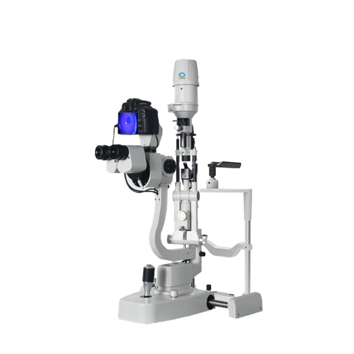
As our new generation of ophthalmic optical coherence tomography imaging system, this system can achieve comprehensive in vivo three-dimensional tomographic imaging of the cornea, iris, retina, and choroid, microvascular non-contrast imaging, and precise measurement of the eye axis. Corneal and retinal microvascular examination can realize the independent output and quantitative analysis of each layer of microvessels. The addition of the axial measurement function makes the equipment more powerful, and accurate measurement provides reliable data for the growth and development of the eyes, distance and nearsightedness. These powerful functions provide an intuitive diagnosis basis for ophthalmic diseases, so as to better understand the pathophysiological mechanism of fundus diseases to assist treatment. It is very helpful to the future gene therapy, stem cell therapy development and new drug research and development.
-
-
-
-
-
-
Feature:
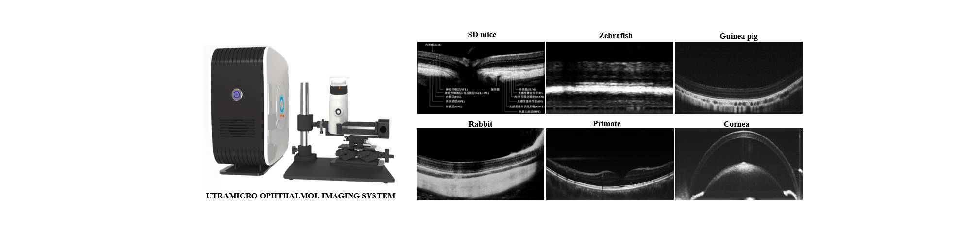
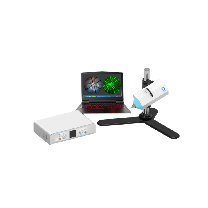
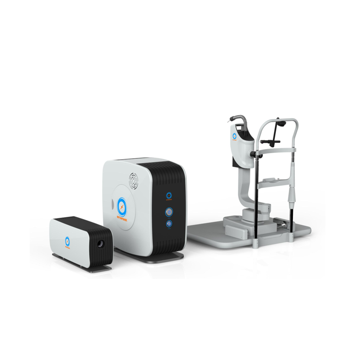
High resolution:Micron level, 1-3mm penetration,imaging of three dimensional tissue in vivo
Free of label:No need for contrast agent, the three-dimensional high resolution microvascular imaging can monitor the pathological changes of various vascular disease models
Fast speed:Fast tomoscan at 350 fps
Wide usage:Microvascular imaging can be performed on various tissues and organs, such as brain tissue, skin, bone ( skull, femoral condyle, surrounding soft tissue, etc. ), viscera ( liver, kidney, uterus, intestine, etc. ), tumors, etc. It can also be applied to a variety of animal models such as human, mouse, zebrafish, quail eggs, etc.